Verhoeff's Van Gieson: A Histological Staining Technique
Verhoeff's Van Gieson is a histological staining technique used to visualize the structural components of tissue samples. It is a combination of two separate staining methods: Verhoeff's stain, which is used to stain elastic fibers, and Van Gieson's stain, which is used to stain collagen fibers. This technique is commonly used in medical research and diagnostic pathology to examine the morphology of tissues and diagnose various diseases. The staining process involves a series of chemical reactions that result in the staining of specific tissue components, allowing researchers to visualize and analyze the tissue structure.
What is the Context of Verhoeff's Van Gieson?
The context of Verhoeff's Van Gieson is rooted in the field of histotechnology, which involves the preparation and staining of tissue samples for microscopic examination. Histological staining techniques like Verhoeff's Van Gieson are essential tools for researchers and pathologists to study the morphology of tissues and diagnose diseases. The technique is particularly useful for examining the structural components of tissues, such as elastic and collagen fibers, which are crucial for understanding various diseases and conditions. For example, Verhoeff's Van Gieson can be used to examine the progression of fibrosis in liver tissue, which is essential for diagnosing and treating liver diseases.
How Does Verhoeff's Van Gieson Work?
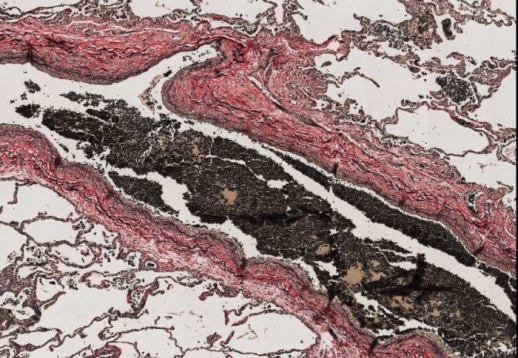
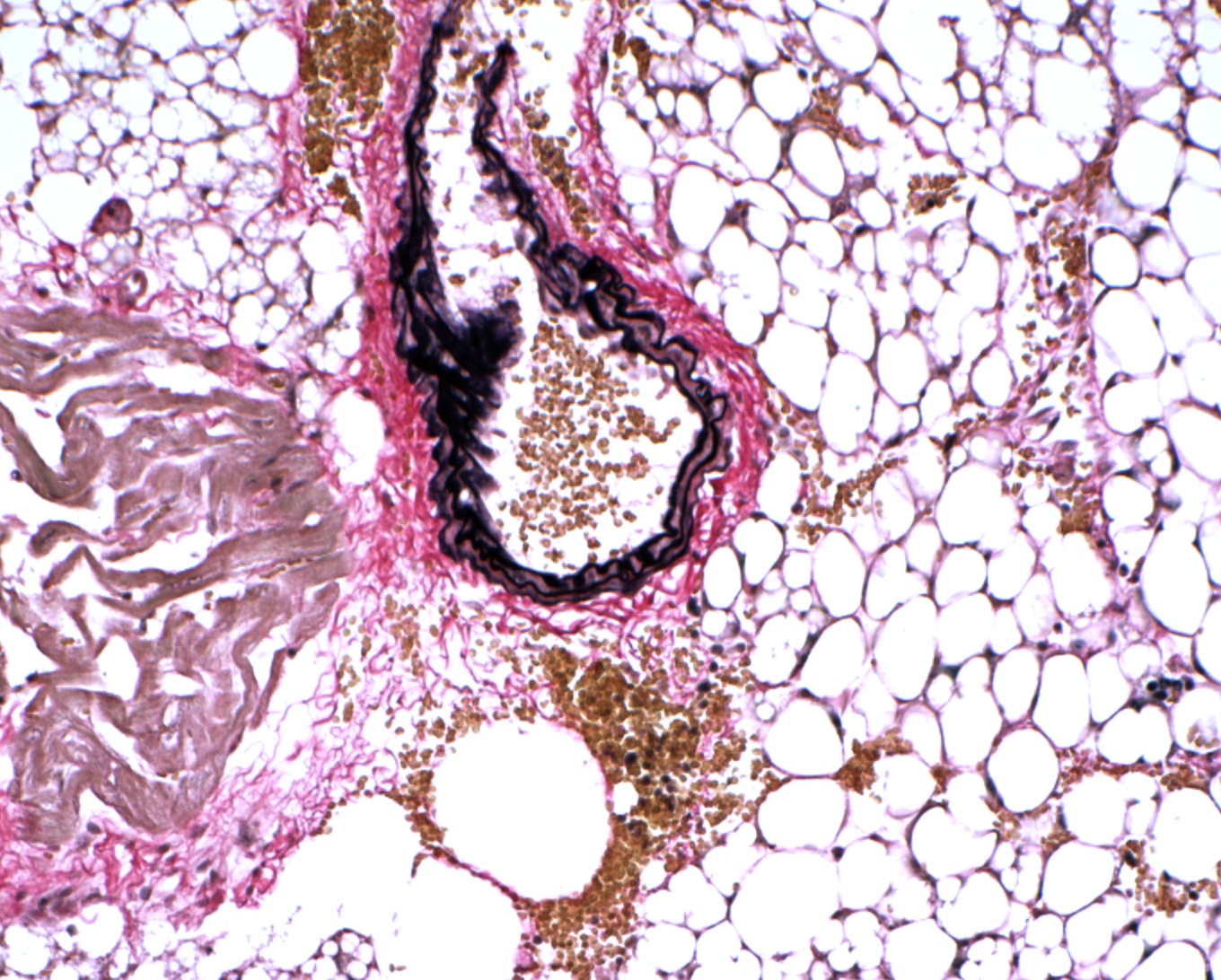
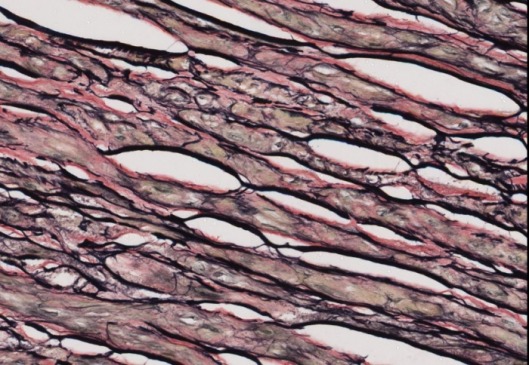
Verhoeff's Van Gieson works by combining two separate staining methods: Verhoeff's stain and Van Gieson's stain. Verhoeff's stain is used to stain elastic fibers, while Van Gieson's stain is used to stain collagen fibers. The staining process involves a series of chemical reactions that result in the staining of specific tissue components. The process begins with the fixation of the tissue sample, followed by the application of the staining solutions. The resulting stained tissue sample can then be examined under a microscope to visualize the structural components of the tissue. For example, the elastic fibers in the tissue sample will appear black, while the collagen fibers will appear red.

What are the Implications of Verhoeff's Van Gieson?
The implications of Verhoeff's Van Gieson are significant in the field of medical research and diagnostic pathology. The technique provides a valuable tool for researchers and pathologists to examine the morphology of tissues and diagnose various diseases. For example, Verhoeff's Van Gieson can be used to diagnose diseases such as liver fibrosis, lung disease, and cardiovascular disease. The technique can also be used to examine the progression of diseases and the effectiveness of treatments. Furthermore, Verhoeff's Van Gieson can be used in combination with other staining techniques, such as Masson's trichrome, to provide a more comprehensive understanding of tissue morphology and disease pathology.
- Verhoeff's Van Gieson is a valuable tool for researchers and pathologists to examine the morphology of tissues and diagnose various diseases.
- The technique provides a detailed visualization of elastic and collagen fibers, which is essential for understanding various diseases and conditions.
- Verhoeff's Van Gieson can be used in combination with other staining techniques to provide a more comprehensive understanding of tissue morphology and disease pathology.
What are the Future Directions of Verhoeff's Van Gieson?
The future directions of Verhoeff's Van Gieson are focused on improving the technique and expanding its applications. Researchers are working to develop new staining methods and protocols that can provide more detailed and accurate visualization of tissue components. Additionally, the technique is being used in combination with other technologies, such as digital microscopy and artificial intelligence, to provide more comprehensive and accurate diagnoses. As the field of histotechnology continues to evolve, Verhoeff's Van Gieson is likely to remain a valuable tool for researchers and pathologists, providing essential information for the diagnosis and treatment of various diseases.
VitroView™ Verhoeff Van Gieson Elastin Stain Kit
VitroView™ Verhoeff Van Gieson Elastin Stain Kit
Verhoeff's Van Gieson - Histotechnology Group DST Project
Verhoeff's Van Gieson - Histotechnology Group DST Project
Verhoeff's Van Gieson Elastic Stain By Hannah Bailey On Prezi
Verhoeff's Van Gieson Elastic Stain by Hannah Bailey on Prezi